Heart Conditions
Angiogram: Everything You Need to Know
4 min read
By Apollo 24/7, Published on - 31 May 2021, Updated on - 18 October 2022
Share this article
0
3 likes

An angiogram, also known as an arteriogram, is an imaging test to examine the blood vessels (arteries or veins) present in the body. The technique of taking an angiogram is called angiography, where an X-ray and a special dye are used to view narrow, blocked, enlarged or malformed blood vessels in any part of the body. An angiogram is performed to check for anomalies in the blood vessels that may be causing issues in the brain, heart, abdomen, kidneys, and even legs.
What are the different types of angiography?
Angiograms can be of different types depending on the organ being examined. Some of the common types of angiograms include:
- Coronary angiograms - for the heart
- Cerebral angiograms - for the brain
- Pulmonary angiograms - for the lungs
- Renal angiograms - for the kidneys
Various techniques are deployed in angiography, some of which include X-rays, Computerised Tomography (CT) and Magnetic Resonance Imaging (MRI) scans. The latter two are better known as CT angiography and MRI angiography.
When is an angiogram advised?
A person is advised an angiogram by their doctor to determine the health and flow of blood through the vessels. Conditions that may require an angiogram to confirm the diagnosis include:
- Peripheral arterial disease - characterised by the low blood supply to the legs
- Angina - pain in the chest due to reduced blood flow to the heart
- Atherosclerosis - narrowing of the blood vessels
- Aneurysm - a bulge in the blood vessel
- Thromboembolism - a blood clot that may block the artery.
Do’s and don’ts before the angiogram
Before the angiography, the doctor would conduct a thorough physical examination followed by a medical history for any known allergies. A proper blood examination may be recommended to determine the ability of the blood to clot. The doctor may provide a few instructions before an angiogram which include:
- Blood-thinning medications such as aspirin or warfarin must be stopped 72 hours before the test.
- Anti-platelet medications such as clopidogrel must be stopped 5 days before the angiogram.
- Short-acting insulin must not be taken on the day of the angiogram. A regular dose of long-acting insulin can be taken.
- The oral anti-diabetic drug, metformin, must not be taken 48 hours before the test.
- One must not eat anything for at least 12 hours before the procedure.
- Clear liquids such as water, tea or clear broth may be taken before the procedure.
What happens in an angiography procedure?
Usually, the person is awake during an angiogram but some patients may require general anaesthesia for the procedure. Here’s what happens in the angiography procedure:
- A long, thin, flexible tube called the catheter is inserted into a large artery in the groin or wrist area by making a small cut in the skin. The site of the cut is numb as the patient is given a local anaesthetic.
- The catheter is carefully guided through the artery until it reaches the area to be examined.
- Once the catheter reaches the target site, a contrast dye is injected through the catheter into the blood vessel.
- This is followed by a series of X-rays that are taken as the dye flows through the blood vessels.
- After the X-rays are taken, the catheter is removed and the cut is closed with a pressure pack to prevent any bleeding. No stitches (sutures) are required.
Do’s and don’ts after the angiogram
After the angiography, the person who has undergone the procedure is taken to the recovery room and made to lie still for a few hours to prevent bleeding from the cut. One must ensure to:
- Take rest for at least 24 hours after the procedure.
- Drink enough water as the contrast dye leaves the body through urine. Thus, drinking enough water would help flush out the dye faster.
- Avoid lifting heavy weights or strenuous exercises for a few days.
- Contact a doctor if one experiences persistent pain, new allergies or swelling at the site of the cut.
Are there any risks associated with angiogram?
Many people experience bruises, soreness or a bump at the site of the cut. However, these problems resolve on their own in a few days. Few people may develop some complications after the procedure. Minor complications of an angiogram include:
- Infection at the site of the cut (characterised by pain, redness, raised temperature and swelling)
- Rashes due to allergy to the dye.
Though rare, major complications that can be seen after an angiogram include:
- Damage to the blood vessel resulting in internal bleeding
- Heart attack
- Stroke
- Kidney damage due to the contrast dye
- Anaphylaxis (serious allergic reaction characterized by difficulty breathing, dizziness and loss of consciousness).
When to contact a doctor
A person must contact their doctor if they experience:
- Bleeding from the cut even after applying pressure for long
- Severe pain that does not resolve after taking medicines
- Pale and cold arm or leg where the cut was made
- Firm and tender lump near the cut.
Takeaway
An angiogram is an extremely important imaging test that helps in determining the source and the extent of the damaged blood vessel. It can help in examining the narrowed or blocked arteries, which if left untreated may result in diseases such as a heart attack, stroke or brain aneurysm. Angiograms help the doctor decide treatment options, which may include angioplasty, bypass surgery or medications, depending on the condition of the person.
Heart Conditions
Leave Comment
Recommended for you

Heart Conditions
Women May Suffer from Heart Diseases Even at a Lower Blood Pressure
A recent study concluded that women could be at higher risk of developing cardiovascular diseases at systolic blood pressure lower than 120 mm Hg.

Heart Conditions
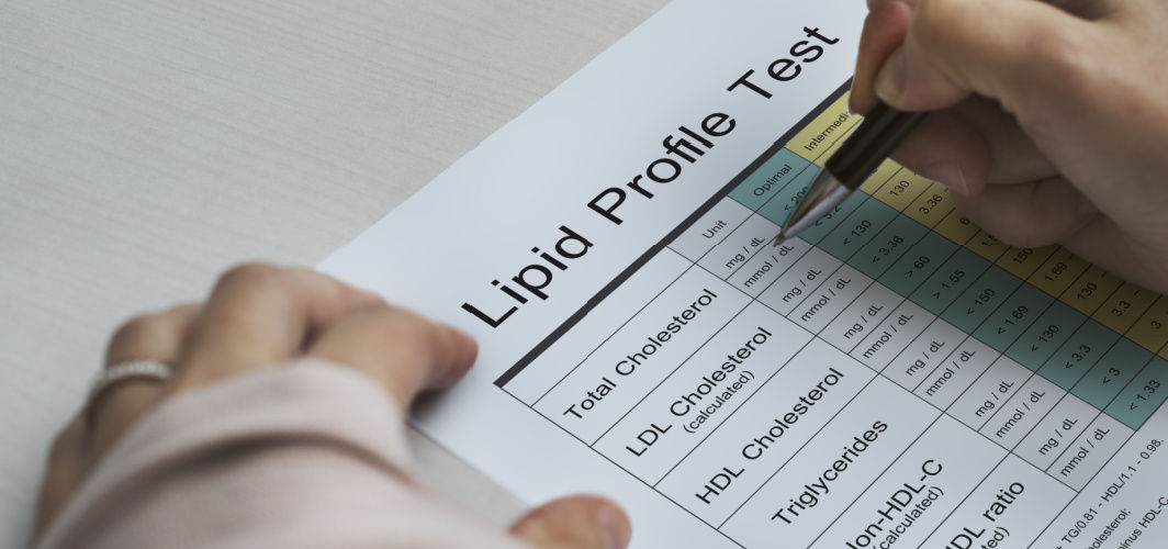
What Does A Lipid Profile Test Indicate?
The lipid profile test plays a crucial role in assessing cardiovascular health. This informative blog covers the importance of lipids in the body, the purpose and timing of the test, preparation guidelines, interpreting the results, and its overall significance for maintaining optimal well-being.
%20(1).jpg?tr=q-80)
Heart Conditions
What is the TMT Test?
The TMT (Treadmill Test) is indispensable for evaluating heart health under stress, offering critical insights into cardiovascular function during physical exertion. TMT test for heart helps diagnose conditions such as Coronary Artery Disease early, determine exercise capacity, and tailor personalised treatment plans to improve heart health.
Subscribe
Sign up for our free Health Library Daily Newsletter
Get doctor-approved health tips, news, and more.
Visual Stories

Lower Your Cholesterol Naturally with These 7 Foods
Tap to continue exploring
Recommended for you

Heart Conditions
Women May Suffer from Heart Diseases Even at a Lower Blood Pressure
A recent study concluded that women could be at higher risk of developing cardiovascular diseases at systolic blood pressure lower than 120 mm Hg.

Heart Conditions
What Does A Lipid Profile Test Indicate?
The lipid profile test plays a crucial role in assessing cardiovascular health. This informative blog covers the importance of lipids in the body, the purpose and timing of the test, preparation guidelines, interpreting the results, and its overall significance for maintaining optimal well-being.
%20(1).jpg?tr=q-80)
Heart Conditions
What is the TMT Test?
The TMT (Treadmill Test) is indispensable for evaluating heart health under stress, offering critical insights into cardiovascular function during physical exertion. TMT test for heart helps diagnose conditions such as Coronary Artery Disease early, determine exercise capacity, and tailor personalised treatment plans to improve heart health.